Original Article
Phacoemulsification in Senile White Mature
Cataracts
Ambreen Gul, Sairam Ahmed, Samana Ali, Ali Raza
DOI 10.36351/pjo.v35i4.881 Pak J Ophthalmol 2019, Vol. 35, No. 4
See end of article for authors affiliations …..……………………….. Correspondence to: Dr. Ambreen Gul Ophthalmology department of Holy family hospital, Lahore Email: amber-gul@hotmail.com …..……………………….. |
Purpose: To evaluate the intraoperative
difficulty, complications and post-operative outcome in patients with white
mature cataracts undergoing phacoemulsification. Study Design: Interventional case series. Place and Duration of Study: Ophthalmology
department of Holy family hospital, from January 2017 to June 2017. Material and Methods: Fifty
Patients who had senile white mature cataract were enrolled in this study. Detailed
preoperative and intraoperative notes were taken. A small capsulorhexis was
attempted initially after staining the capsule with trypan blue. An initial
cut was made with cystotome and it was enlarged with utrata forceps.
Phacoemulsification was done with stop and chop technique. Intraoperative
difficulties related to continuous curvilinear capsulorhexis,
phacoemulsification and post-operative visual outcomes were analyzed. Post-operative
examinations were done at day 1, 1 week, 1 month and 3rd month. Results: There were 28 males (56%) and
22 females (44%). The mean age was 63.18 ± 7.997. The mean preoperative best
corrected visual acuity (BCVA) was 0.0276 ± 0.013 with Snellen chart, (0.01-0.05)
and mean post-operative BCVA was 0.638 ± 0.305 (0.1-1.0). Mean phaco time was
4.08 ± 1.03 minutes (2.08-6.66). Posterior capsular rupture occurred in 3
(6%) and vitreous loss occurred in 1 (2%) patient. 5 (10%) cases were
converted to extracapsular cataract extraction (ECCE). Postoperatively, 10
(20%) patients had transient corneal edema, 3 patients (6%) had persistent
corneal edema and corneal burn treated with steroids and hyper osmotic
agents. Conclusion: White mature cataract is a
challenge for cataract surgeon, yet by means of additional dyes and proper
techniques and expertise, the rate of complications during
phacoemulsification can be reduced. Key
Words: Phacoemulsification, Cataract, Trypan blue, Posterior capsular
rupture, Corneal edema. |
Cataract is the most frequent
treatable blindness worldwide. In developing countries, white mature cataracts
are seen very frequently1. In Pakistan, the most common cause of
blindness and low vision is an un-operated cataract. A review conducted in
Pakistan yielded prevalence of 1.78% and found out that cataract is a major
cause of treatable blindness constituting 66.7% of total blindness. Mature and hypermature
cataracts constitute a major volume of surgical load2.
Surgical removal of white mature cataracts
presents special challenges to the surgeon. It is associated with high rate of
intraoperative and postoperative complications; such as incomplete CCC, radial
tears in anterior capsule extending to equator and posterior capsule, rupture
of posterior capsule, vitreous loss, nucleus drop, IOL dislocation, corneal
burns, IOP rise, persistent corneal edema and anterior chamber reaction3-5.
Continuous curvilinear capsulorhexis and emulsification of hard nucleus are two
critical steps that make phacoemulsification challenging in these cases. Visualization
of the anterior capsule depends on red reflex, which is compromised in eyes
with white mature cataract. The capsule is extra fragile and seepage of
liquefied cortical material causes the capsulorhexis tear to extend to the
periphery on account of high intracapsular pressure. The anterior capsule may
undergo disintegration with deposition of calcium or growth of focal plaques
may hinder the capsulorhexis6.
The use of Trypan blue facilitates CCC
formation, provides a safe surgery, resulting in decrease in intraoperative
complications. Nuclei of varying hardness may be masked by a totally opaque
cortex. After the nucleus is removed by the divide and conquer or the phaco
chop technique, a posterior chamber intraocular lens can be implanted. Even for
an experienced surgeon harder nucleus will require a longer time and higher
power of phacoemulsification. A plaque or residual posterior capsule is
observed in spite of successful surgery7.
This study was conducted
in the department of ophthalmology, Holy family hospital, Rawalpindi. We
evaluated the safety and postoperative visual outcome in patients undergoing
phacoemulsification in white mature cataract.
MATERIAL AND METHODS
After approval from the local ethical committee,
this cross sectional study was carried out between January 2017 and June 2017. In
this study, 50 eyes of the 50 patients with white mature cataract were
evaluated prospectively. All eyes in mature cataract group lacked red fundus
reflex. Cataracts appearing white on slit lamp examination were defined as
white mature, hypermature or brunescent cataracts. Patients with diabetes
mellitus, glaucoma, pseudo exfoliation, complicated cataract, poor pupil
dilation (< 5mm), history of ocular surgery, laser treatment or trauma were
excluded. Written informed consent was obtained from each patient.
Preoperative ocular examinations included Snellen
visual acuity, detailed biomicroscopic examination including anterior chamber
examination, Goldmann Applanation tonometry, axial length and anterior chamber
depth measurements with A-scan ultrasonography. Keratometry was performed using
an Autokeratorefractometer.
Mydriacyl 1% and phenylephrine 2.5% eye
drops were used for mydriasis, 1 hour before the surgery. Three surgeons
performed all surgeries. Endocapsular phacoemulsification was performed in all
cases by using infinity Alcon and Opticon phacoemulsification unit by one of
the three surgeons in an identical manner. Topical or retrobulbar anaesthesia
was used. Pieces of cotton sponge impregnated with proparacaine HCL 0.5% were
placed deep into superior and inferior fornix, for 15 minutes before surgery
for topical anaesthesia. Retro bulbar injection was performed using a 23-gauge
needle, 3 ml of 2% lidocaine was given intraconallly.
Nuclear hardness was subjectively evaluated
by the surgeon intraoperatively during phacoemulsification and was graded as soft,
semi soft, medium, hard, very hard. Effective phaco time displayed by the phacoemulsification
unit for each surgical procedure was recorded.
A three-step clear corneal tunnel incision
was made with a 3.2 mm disposable metal blade and a side port incision was made
with side port knife. Staining of the anterior capsule was done with trypan
blue under air. After injection of dispersive viscoelastic sodium chondroitin
sulfate-sodium hyaluronate into the anterior chamber of eyes, continuous
curvilinear capsulorhexis was performed. Before completing the CCC liquefied
milky cortex was aspirated in eyes to decrease high intracapsular pressure for the
safety of capsulorhexis. Radial tears occurred in patients and conversion to
extra capsular cataract extraction was preferred in these patients. After CCC hydro
dissection was performed carefully in these cases because posterior capsule is
thinner and more fragile in hard cataracts. Nucleus was removed by using divide
and conquer or stop and chop to prevent damage to corneal endothelial cells as
more energy is used in hard cataracts. Cortex was aspirated with irrigation and
aspiration and anterior chamber was filled with cohesive viscoelastic substance
and foldable monofocal posterior chamber IOL was implanted in the capsular bag
through an injector system. The viscoelastic material was aspirated completely,
the entrances were closed with stromal hydration and for endophthalmitis
prophylaxis sub-conjunctival antibiotic ceftriaxone and steroid dexamethasone
injection was given.
Post-operative examinations were done at 1
day, 1 week, 1st and 3rd months. After surgery patient
used topical antibiotics 2 hourly, steroid 4 hourly, daily for 1 week and
topical steroid was tapered for subsequent 4 weeks. Patients who had transient
or persistent corneal edema were managed with intense topical steroid and hyper-osmotic
agents that took 6 weeks to resolve. Preoperative and postoperative BCVA values
were used for statistical analyses. Preoperative and intraoperative findings as
well as postoperative outcomes were analyzed.
SPSS version 21 was used
for statistical analysis. Data was compared by using paired t test.P< 0.05
was accepted as significant.
RESULTS
Out of total 50 patients, there were 28 males
(56%) and 22 females (44%).The mean age was 63.18 ± 7.997 years with a range of
48 to 78 years. Out of 50 patients, 22 (44%) patients had mature cataract, 15
(30%) patients had hyper-mature cataract, 13 (26%) patients had Brunescent
cataract. Pre-operative best-corrected visual acuity (BCVA) was recorded which
ranged from light perception (0.01) to 3/60 (0.05) (Table 1). Forty one patients
had uneventful surgery with no intraoperative complications. Intraoperative
complications included premature entry of the tunnel into the anterior chamber,
incomplete capsulorhexis, and posterior capsular tear, conversion to a manual
non-phacoemulsification technique, intraoperative miosis, and iris chafing.
Intraoperatively Trypan blue staining was used in all patients. Aspiration of
liquefied milky cortex was performed in patients before completing CCC. Radial
tears occurred in 5 (10%), Posterior capsular rupture occurred in 3 (6%),
vitreous loss occurred in 1 (2%) patient. 5 (10%) cases were converted to extra
capsular cataract extraction (ECCE) with IOL implantation in the sulcus. No
nucleus drop occurred.
Mean phacoemulsification time was 4.08±1.03
(SD) minutes with a range of 2.08-6.66 minutes (Figure 1). The mean
preoperative intraocular pressure IOP was 15.90 ± 1.799 (SD) mmHg range (12-22)
and mean post op IOP was 11.98 ± 2.035 (SD) mmHg range (10 – 18). The mean post
operative IOP was significantly lower than that of preoperative value.
Preoperatively Phacomorphic glaucoma was present in none patient and their IOP
were significantly lower postoperatively without any medication.
Postoperatively 10 (20%) patients had
transient corneal edema lasting one week which resolved with topical steroid
therapy, 3 patients (6%) had persistent corneal edema and corneal burn treated
with intense topical steroids and hyper osmotic agents, their corneal edema
resolved within 6 weeks. Corneal burn occurred in two (4%) cases. Severe
striate keratitis occurred in four (8%) patients. three (6%) patients had three
plus cell count in anterior chamber, which resolved with intense topical
steroids in 1 month. Postoperatively IOL dislocation occurred in one (2%)
patients. Postoperative IOP rise occurred in none of the cases. These postoperative
complications are summarized in table 2.
At 3rd post-operative month, BCVA ranged from
Table 1: Pre- operative best corrected
visual acuity.
Grading of Visual Acuity (Snellen
Decimal Fraction) |
Frequency |
Percent |
Valid
Percent |
Cumulative
Percent |
Perception of light(0.01) |
15 |
30.0 |
30.0 |
30.0 |
Hands Movement(0.02) |
26 |
52.0 |
52.0 |
82.0 |
Counting finger up to 3 meters(0.03) |
6 |
12.0 |
12.0 |
94.0 |
Counting finger better than 3 meters
to 6/60(0.04-0.05) |
3 |
6.0 |
6.0 |
100.0 |
Total |
50 |
100.0 |
100.0 |
|
Table 2: Postoperative complications.
Post-operative Complications |
Frequency |
Percent |
Valid
Percent |
Cumulative
Percent |
No complication |
29 |
58.0 |
58.0 |
58.0 |
Transient Corneal Edema |
10 |
20.0 |
20.0 |
78.0 |
Persistent Corneal Edema |
1 |
2.0 |
2.0 |
80.0 |
Corneal Burn |
1 |
2.0 |
2.0 |
82.0 |
Striate Keratitis |
4 |
8.0 |
8.0 |
90.0 |
Post-operative uveitis |
3 |
6.0 |
6.0 |
96.0 |
Corneal burn plus striate keratitis |
1 |
2.0 |
2.0 |
98.0 |
IOL dislocation plus persistent
corneal edema |
1 |
2.0 |
2.0 |
100.0 |
Total |
50 |
100.0 |
100.0 |
|
Table 3: Post-operative best corrected
visual acuity.
Grading of Visual Acuity (Snellen
Decimal Fraction) |
Frequency |
Percent |
Valid
Percent |
Cumulative
Percent |
less than or equal to 6/60 (≤ 0.1) |
2 |
4.0 |
4.0 |
4.0 |
6/36 to 6/18 (0.1-0.3) |
10 |
20.0 |
20.0 |
24.0 |
6/12 to 6/6 (0.5-1.0) |
38 |
76.0 |
76.0 |
100.0 |
Total |
50 |
100.0 |
100.0 |
|
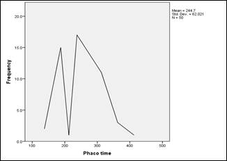
Fig. 1: Mean Phacoemulsification time
≤6/60 (≤
0.1) to 6/6 (1.0) (Table 3). BCVA was significantly higher than mean preoperative
BCVA.
(P < 0.001).
DISCUSSION
Mature and hypermature cataracts constitute
a significant volume of the cataract surgical load in ophthalmic practice in
the developing countries. There were an estimated 1,140,000 (962,000-1,330,000)
blind adults in Pakistan in 2003. Countryside areas had a higher frequency of
blindness than did urban areas (3.8% vs. 2.5%). Most patients have advanced
stages of cataract with intumescent, mature or hypermature cataract. Majority
of these patients are less privileged8.
White mature cataracts are a challenge for
cataract surgeon and carry some difficulties. The most critical step of phacoemulsification
surgery is Continuous Curvilinear Capsulorhexis. If it is not complete, some
intraoperative complications such as posterior capsule rupture, vitreous loss
and nucleus drop may occur. Because the red reflex is compromised in white
cataract, it is difficult to complete CCC safely. Trypan blue provides a safe
CCC7,9.
General recommendations for visualization
of anterior capsule in eyes with mature white cataract include dimming the
operation room lights, increasing the magnification of microscope, using
oblique illumination, capsule dyes. Giammaria D et al and Wong et al, stained
the capsule under an air bubble, it was reported that using the dye under the
dispersive viscoelastic material was easier and safe. The air bubble technique
was reported to be time consuming7,10. The rate of conversion to
ECCE in white cataracts as a result of an incomplete CCC has been as low as 3.85%
when Trypan blue is used compared to 28.3% when no staining was used. In our
study, we used Trypan blue in all patients, and radial tears occurred in five
patients and rate of conversion to ECCE was 10%. It has been reported that Trypan
blue did not cause any inflammation, corneal edema, corneal thickening, decrease
in endothelial cell count and IOP rise11. Portes et al demonstrated
that Trypan blue caused lens epithelial cell death, which supported the
hypothesis that staining with Trypan blue 0.1% helps reducing the incidence of
posterior capsule opacification after cataract surgery. The frequency of
capsular rupture and vitreous loss can be reduced by staining the anterior
capsule with the Trypan blue to identify the capsular tear at an early stage.
We achieved a 5 mm capsulorhexis in most of the cases 12.
Kara junior et al recommended the mini
rhexis technique for white intumescent cataracts in which primarily a small CCC
was performed then enlarged. Two stages CCC prevented unanticipated radial
tears of the initial capsulotomy due to elevated intra capsular pressure13.
We aspirated liquefied milky cortical matter via cannula in 15 patients before
finishing CCC to avoid sudden radial tears due to highly intracapsular
pressure. Chen and Wu suggested automated irrigation and aspiration by lowering
of BSS bottle to aspirate the liquefied milky lens contents before
phacomulsification14. Daglioglu et al suggested an innovative
capsulorhexis technique in white cataract surgery in which CCC was completed by
using an irrigation and aspiration system by phaco machine, it was found safe
in white cataracts15.
Although hydro dissection was not
recommended in white mature cataracts, we observed that gentle hydro dissection
broke the cortico-capsular adhesions that could resist free nucleus rotation1.
Singh et al reported cortico-capsular adhesions resulted in different nucleus
rotation in brunescent and black cataracts. Nucleus rotation is critical for
phacoemulsification16.
Posterior capsule is not only weak but also
flaccid with wrinkles and laxity that makes it prone to be ruptured. The
problem is worsened by absence of any epinucleus that protects the posterior
capsule. A useful step is to inject a dispersive non–cohesive viscoelastic
behind the nucleus during the phacoemulsification, which will provide an
artificial epinucleus to keep the posterior capsule back from the operating
plane and stabilize the nucleus against tumbling17.
In Brunescent and black cataracts, the lens
fibers were found to be very cohesive thus making division difficult. White
cataracts in our study were usually brittle and not very hard; they were safely
divided and emulsified. During the division and aspiration of the nucleus, edge
of the hard nucleus may cut the posterior capsule, resulting in rupture; also
radial tears in anterior capsulotomy may extend to posterior capsule and cause
rupture. Therefore, the incidence of posterior capsule rupture is higher in
mature cataracts18. In our study rate of posterior capsular rupture
was 6%.
Phacoemulsification of hard nucleus
requires higher ultrasonic energy, which is partially converted to heat energy
causing corneal endothelial damage and corneal burns. Fluid dynamics during
phacoemulsification may cause endothelial cell damage if it lasts longer18.
With aging, endothelial cell count decreases, this is another risk for patients
with mature cataracts; therefore, chances of post-operative corneal edema is
higher in patients with mature cataract18,19.
In our study, we did not encounter
complications of capsular fibrosis and geometrical decentration. In another study,
capsular fibrosis was reported to occur in 12% of eyes with white mature
cataracts all of which had a capsulorhexis diameter of less than 5 mm20.
Small capsulorhexis leads to capsule contraction.
Yuan et al recommended that ophthalmic
viscosurgical device assisted sutureless cataract surgery, usually without
additional instruments, or sutures presented an efficient and uncomplicated
technique for managing a brunescent or mature cataract21. Venkatesh
et al compared manual small incision cataract surgery with phacoemulsification
for white cataract and reported that both techniques achieved excellent visual
outcomes with low complication rates22. Wong et al suggested that
micro-incisional cataract surgery with bimanual phacoemulsification appeared to
be a hopeful alternative for management of white cataracts23. Kim
and Jang proposed drill and chop technique for hard cataracts, which required
complete engagement of central nucleus by phaco tip. First, a hole was drilled
into the endonucleus by rotating the Kelman phaco tip clockwise, nucleus was
deeply impaled horizontally and completely engaged by phaco tip followed by
vertical chopping and it resulted safer and more effective vertical chopping in
patients with harder cataracts24. Li et al described the peripheral
radial chop technique in phacoemulsification of harder nuclei and stated that
it was effective without grave complications in hands of skilled surgeons25.
The limitation of our study was that it was performed in one
center. More studies need to be performed with larger number of patients in
multiple centers.
CONCLUSION
White mature cataract is
a challenge for phaco surgeons but with appropriate techniques such as two
stage capsulorhexis and use of additional capsule staining dyes can achieve
excellent visual outcomes and low complication rates.
REFERENCES
1.
Ermisş SS, Oztürk F, Inan UU. Comparing the efficacy and safety of phacoemulsification in white
mature and other types of senile cataracts. Br J Ophthalmol. 2003 Nov;
87(11):1356-9.
2.
Khan AQ, Qureshi B, Khan D. Rapid assessment of cataract blindness in age 40 years and above
in district skardu, Baltistan, Northern areas, Pakistan. Pak J Ophthalmol.
2003; 19: 84-9.
3.
Ilavska M, Kardos L.
Phacoemulsification of mature and hard nuclear cataracts. Bratisl Lek Listy, 2010;
111 (2): 93-6.
4.
Susic N, Brajkovic J, Susic E, Kalauz-Surac I. Phacoemulsification in eyes with white cataract. Acta Clin Croat, 2010;
49 (3): 343-5.
5.
Hawlina M, Stunf S, Hvala A. Ultrastructure of anterior lens capsule of intumescent White
cataract. Acta Ophthalmol. 2011; 89 (4): e367-70.
6.
LoBue SA, Tailor P, LoBue TD. A Simple, Novel Approach to Capsulorhexis Formation in the Setting
of A Mature Cataract and Miotic Pupil. Clin Ophthalmol. 2019 Dec 2; 13: 2361-2367.
7.
Giammaria D, Gianotti M, Scopelliti A, Pellegrini G, Gianotti B. Under-air staining of the anterior capsule using trypan blue with
a 30 G needle. Clin Ophthalmol. 2013; 7: 233-235.
8.
Jadoon MZ, Dineen B, Bourne RR, Shah SP, Khan MA, Johnson GJ, et
al. Prevalence of blindness and
visual impairment in Pakistan: The Pakistan National Blindness and Visual
Impairment Survey. Invest Ophthalmol. 2006; 11: 4749-55.
9.
Rossiter J, Morris A.
Trypan blue vital staining of the anterior lens capsule in the management of
cataract in true exfoliation of the lens capsule. Eye, 2005; 19: 809-10.
10.
Wong VW, Lai TY, Lee GK, Lam PT, Lam DS. A prospective study on trypan blue capsule staining under air vs
under viscoelastic. Eye (Lond.), 2006; 20 (7): 820-825.
11.
Cheour M, Ben Biahim F, Zauad A. Trypan blue capsule staining for phacoemulsification in white
cataract. J Fr Ophthalmol. 2007; 30 (9): 914-917.
12.
Portes AL, Almeida AC, Allodi S, Monteiro ML, Miguel NC. Trypan Blue Staining for Capsulorhexis; ultrastructural effect on
lens epithelial cells and capsules. J Cataract Refract Surg. 2010; 36 (4) 582-7.
13.
Kara-Junior N, de Santhiago MR, Kawakami A, Caricondo P, Hidaet
WT. Mini-rhexis For White
Intumescent Cataracts. Clinics (sao Paulo). 2009; 64 (4): 309-312.
14.
Chen YJ, Wu PC.
Automated irrigation/aspiration before phacoemulsification in eyes with white
cataracts. Ophthalmic Surg Lasers Imaging, 2005; 36 (2): 118-21.
15.
Daglioglue MC, Coskun M, Ilhan O, Tuzco EA, Ilhan N, Ayintap E, et
al. A novel capsulorhexis
technique in white cataract surgery. Semin Ophthalmol. 2014; 30 (4): 264-267.
16.
Singh R, Vasavada A, Janaswamy G. Phacoemulsification of Brunescent and black cataracts. J Cataract
Refract Surg. 2001; 27: 1762-9.
17.
Cetinkaya S, Gurdag T, Akcam N. Phacoemulsification in eyes with white mature cataract. Sch. J. App.
Med. Sci. 2015; 3 (2c): 701-4.
18.
Bilgin B, Eltutar K, Sezgin BI. Comparison of phacoemulsification results of mature and
nucleocortical cataracts. Turk J Ophthalmol. 2006; 36: 219-22.
19.
Sizmaz S, Peli A, Yaycioglu RA. The use of trypan blue in patients with white cataract. Turk J
Ophthalmol. 2007; 37: 178-181.
20.
Shahid E, Sheikh A, Fasih U. Complications of hypermature cataract and its visual outcome. Pak
J Ophthalmol. 2011; 27 (2): 58-62.
21.
Yuan X, Song H, Hua X. Ophthalmic viscosurgical device- assisted sutureless-incision
cataract surgery for a hard nucleus or mature cataract. J Cataract Refract
Surg. 2014; 40 (4): 517-20.
22.
Venkatesh R, Tan CS, Sengupta S, Ravindran RD, Krishan KT, Chang
DF. Phacoemulsification versus
manual small incision cataract surgery for white cataract. J Cataract Refract
Surg. 2010; 36 (11): 1849-54.
23.
Wong VW, Lai TY, Lee GK. Safety and efficacy of micro-incisional cataract surgery with
bimanual phacoemulsification for white mature cataract. Ophthalmologica. 2007; 221
(1): 24-8.
24.
Kim DY, Jang JH.
Drill and chop: modified vertical chop technique for hard cataract. Ophthalmic
Surg Lasers Imaging, 2012; 43 (2): 169-72.
25.
Li SW, Xie LX, Song ZH. Peripheral radial chop technique for phacoemulsification of hard
cataracts. Chin Med J (Engl.). 2007; 120 (4): 284-86.
Author’s Affiliation
Dr. Ambreen Gul
Senior Registrar
Department of Ophthalmology,
Holy family hospital, Rawalpindi medical university, Rawalpindi
Dr. Sairam Ahmed
Resident
Department of
Ophthalmology, Holy family hospital, Rawalpindi Medical University, Rawalpindi
Dr. Samana Ali
House Surgeon
Department of
Ophthalmology, Holy Family Hospital, Rawalpindi Medical University, Rawalpindi
Dr. Ali Raza
Professor and Head
of Department
Department of Ophthalmology, Holy Family Hospital,
Rawalpindi Medical University, Rawalpindi
Author’s Contribution
Dr. Ambreen Gul
Study design, data collection,
analysis, manuscript writing and final review.
Dr. Sairam Ahmed
Data analysis.
Dr. Samana Ali
Data collection and analysis.
Dr. Ali Raza
For putting intellectual input and
supervising the study.